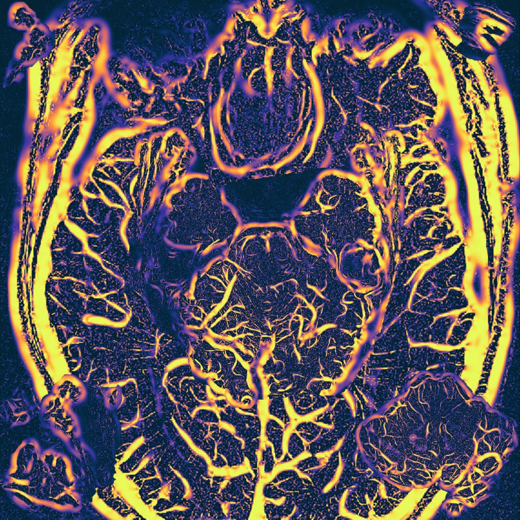
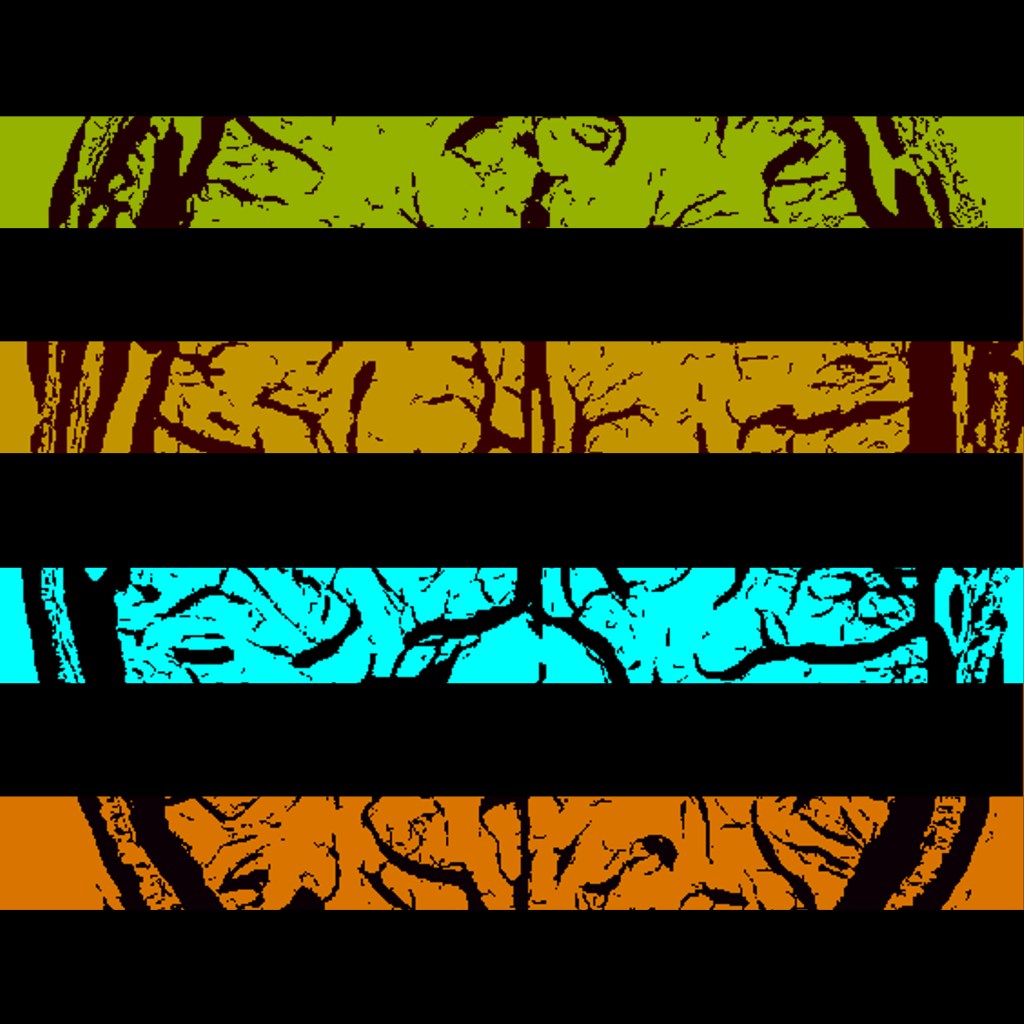
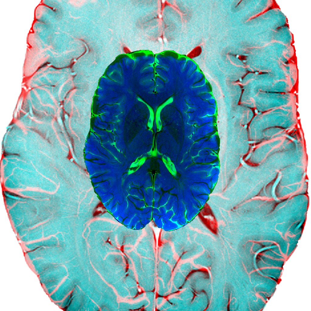
2019 Ultrahigh Field Imaging

This image was made by doing an edge detection of a high-resolution structural image. The edge detection gradient was amplified, and the resulting slices were mirrored.
An edge (detection) gradient, mirrored
Susceptibility weighted images produce amazing visualization of the vessels. A Frangi filter was used to emphasis the  vessels, and elements of the brain were hidden within.
The brain is in the mind… or is the mind in the brain?
The edge detection filter was used to focus on the area around the cerebellum and the gradient was emphasized.
The edge gradient makes the tree of life shine
Yael Jacob used fMRI to produce a tinker toy representation and we arranged the results to produce this image.
Tinker toy representation of fMRI
A slice of a whale brain, both human like and... not.
A whale (brain) of a 4th of July
A SWI image put through a vessel detection algorithm, and sliced.
Black out blinds
an SWI image overlaid on a T2TSE image
Tripping
Diffusion tracts emanating from the hippocampus, used as a stamp to create a Christmas  collage.
Diffuse Christmas

Leave a Reply

Fill in your details below or click an icon to log in:

WordPress.com Logo

You are commenting using your WordPress.com account. Log Out /  Change )

Facebook photo

You are commenting using your Facebook account. Log Out /  Change )

Connecting to %s